

In cardiovascular scenarios, the interpretation of an electrocardiogram (ECG) is frequently the primary diagnostic pivot point upon which clinical decision-making rests. Utilizing pristine, artifact-free, high-fidelity ECG waveforms ensures that learners are evaluated strictly on their diagnostic acumen rather than their ability to guess through visual clutter.
When designing simulation curricula, selecting the appropriate dysrhythmia requires a precise alignment with the learners’ training level. Introducing advanced electrophysiological concepts to novice students can induce cognitive overload, while presenting baseline rhythms to expert providers fails to challenge critical clinical thinking.
The following analysis examines four critical, hard-to-source ECG presentations – outlining their electrophysiological mechanics, target learner levels, and strategic deployment methods within simulation scenarios to optimize pedagogical impact.
1. AV Block 2nd Degree, Mobitz II
Download this waveform via theSimTech ECG/EKG Images Database.
Electrophysiological Overview
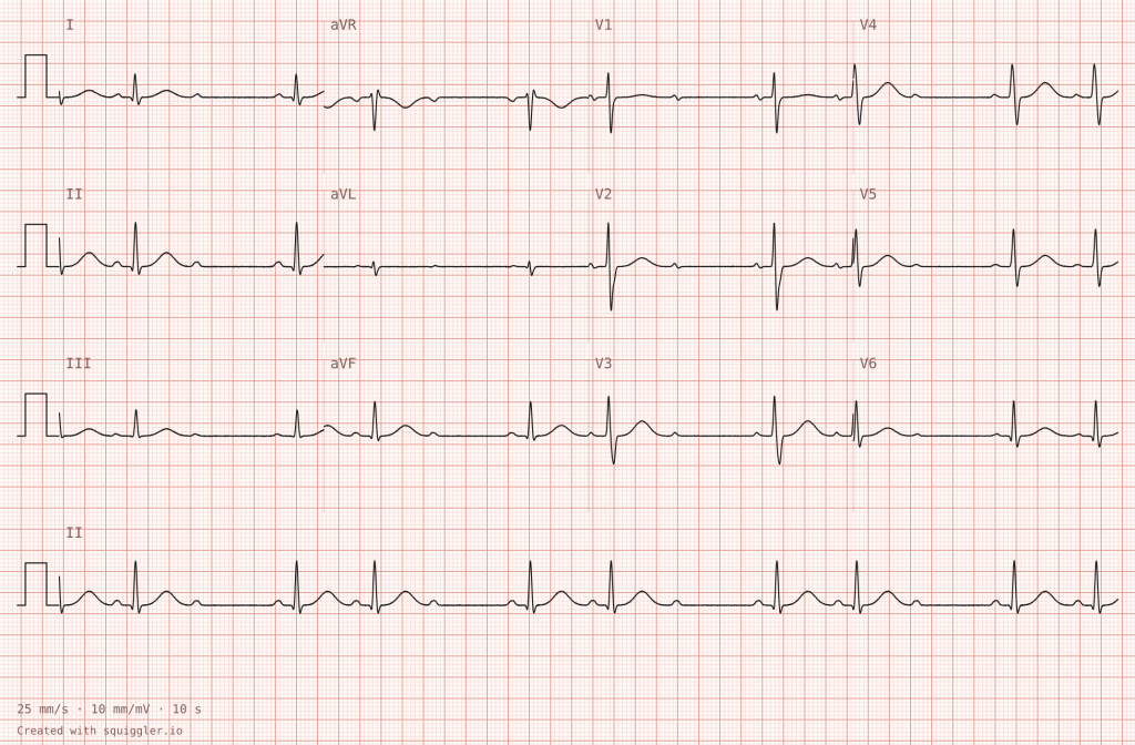
Mobitz II second-degree atrioventricular (AV) block is characterized by an intermittent, unexpected failure of conduction through the Bundle of His or bundle branches. Unlike Mobitz I (Wenckebach), where the PR interval progressively elongates until a QRS complex is dropped, a Mobitz II block maintains a constant, unvarying PR interval across all conducted beats immediately preceding the non-conducted P wave. Because this block occurs below the AV node, it carries an exceptionally high risk of sudden degeneration into complete (third-degree) heart block, severe bradycardia, and hemodynamic collapse.
Target Learner Cohort
Intermediate to Advanced: Paramedic students, Emergency Medicine residents, and Critical Care/Emergency nursing staff.
Pedagogical Strategy
Simplifying the Concept
Utilize this waveform to demonstrate baseline consistency contrasted with sudden conduction failure. The clear presentation allows instructors to highlight the stability of the sinus node (manifested by regular P-to-P intervals) alongside an unpredictable infra-nodal block.
This serves as an ideal clinical model for mapping the anatomical pathways of the cardiac conduction system.
Challenging the Learner
Embed this rhythm into an active emergency scenario. Initiate the simulation with a patient presenting with non-specific symptoms such as transient dizziness or weakness. Once the learner obtains the ECG, introduce a sudden acute drop in blood pressure or a brief syncopal episode. Instructors can evaluate the learner’s readiness to initiate transcutaneous pacing or prepare for temporary transvenous pacing, demanding decisive intervention before the rhythm degenerates into a lethal asystolic pause.
2. AV Block 2:1
Download this waveform via theSimTech ECG/EKG Images Database.
Electrophysiological Overview
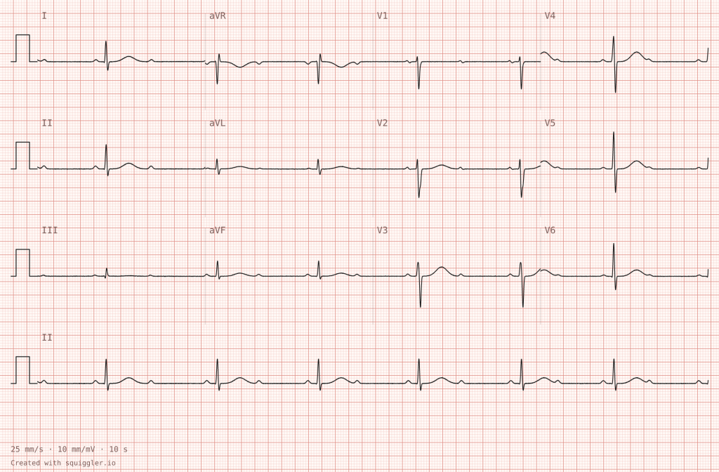
A 2:1 AV block occurs when every second P wave fails to conduct to the ventricles, resulting in a regular ventricular rate that is exactly half the atrial rate. Diagnostically, a 2:1 block occupies a unique gray area: because there are no two consecutive conducted QRS complexes, it is impossible to observe a progressive lengthening of the PR interval (Mobitz I) or a fixed PR interval across multiple beats (Mobitz II). Differentiating the underlying mechanism requires analyzing the width of the conducted QRS complex, examining long rhythm strips for subtle changes, or evaluating the patient’s response to autonomic interventions.
Target Learner Cohort
Advanced and Specialist: Cardiology fellows, Advanced Cardiac Life Support (ACLS) instructors, and Senior Emergency department providers.
Pedagogical Strategy
Simplifying the Concept
The regular, alternating pattern of a 2:1 block provides a structured template for teaching meticulous telemetry analysis. Educators can use this rhythm to train students in the systematic application of calipers to verify absolute atrial regularity (P-P intervals) alongside a perfectly matched, yet bradycardic, ventricular output.
Challenging the Learner
Use this ambiguous presentation to test diagnostic nuance and pathophysiology deduction. Challenge advanced learners to determine the structural level of the block based on indirect visual clues—such as a wide QRS complex implying an infranodal Mobitz II origin, versus a narrow QRS complex suggesting a nodal Mobitz I origin. Introduce a clinical dilemma where the patient is border-line unstable; the learner must defend their therapeutic choices, recognizing that administering atropine could inadvertently worsen an infranodal Mobitz II block by accelerating the atrial rate.
3. Ventricular Paced Rhythm
Download this waveform via theSimTech ECG/EKG Images Database.
Electrophysiological Overview
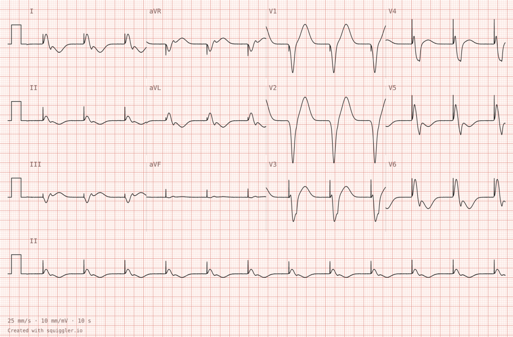
An artificial ventricular pacemaker stimulates the ventricular myocardium directly via an electrode implanted in the right ventricle. On a standard 12-lead ECG, this produces a sharp, narrow vertical deflection—a pacing spike—immediately preceding the QRS complex. Because ventricular depolarization originates from the apex of the right ventricle rather than the normal His-Purkinje system, sequential activation of the ventricles is disrupted. This results in a wide, distorted QRS complex exceeding 120 milliseconds, typically exhibiting a left bundle branch block (LBBB) morphology with secondary ST-segment and T-wave discordance.
Target Learner Cohort
Novice to Intermediate: Undergraduate nursing students, Emergency Medical Technicians (EMTs), and foundational telemetry technicians.
Pedagogical Strategy
Simplifying the Concept
The distinct separation between the artificial pacing spike and the subsequent wide QRS complex provides an ideal teaching baseline. It allows novice learners to easily differentiate between mechanical device intervention and intrinsic cardiac electrical activity, solidifying their understanding of ventricular depolarization pathways.
Challenging the Learner
Deploy this rhythm to reinforce the critical concept of electrical versus mechanical capture. Present a scenario where the patient is profoundly bradycardic and obtunded, yet the cardiac monitor displays a regular ventricular paced rhythm. The learner must look beyond the screen to physically verify a corresponding carotid or femoral pulse.
For advanced learners, this rhythm can be used to teach the identification of acute myocardial infarction hidden within a paced morphology, requiring the application of specialized criteria (such as Sgarbossa or modified Smith-Sgarbossa criteria) to diagnose ischemia.
4. Pre-Excited Atrial Fibrillation
Download this waveform via theSimTech ECG/EKG Images Database.
Electrophysiological Overview
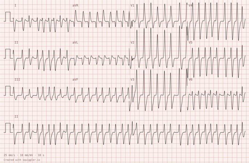
Pre-excited Atrial Fibrillation occurs when a patient with an accessory pathway – such as the Bundle of Kent in Wolff-Parkinson-White (WPW) syndrome – develops atrial fibrillation. In standard atrial fibrillation, the AV node acts as a gatekeeper, slowing down the chaotic atrial impulses. However, an accessory pathway lacks this decremental conduction capability. Impulses travel down both the normal AV node and the fast accessory tract simultaneously, leading to an exceptionally rapid ventricular response that frequently exceeds 200 to 250 beats per minute. The resulting ECG is characterized by a triad of features: it is fast, broad (due to varying degrees of delta-wave fusion), and irregularly irregular.
Target Learner Cohort
Advanced, Critical Care, and Emergency Specialties: Flight Paramedics, Critical Care Physicians, and Intensive Care Unit resuscitation teams.
Pedagogical Strategy
Simplifying the Concept
Real-world examples of pre-excited AFib often appear as a chaotic blur. Presenting a pristine rendering allows educators to break down the diagnostic features systematically. Instructors can guide learners to observe how the QRS morphology dynamically changes from beat to beat based on the fluctuating competition between the AV node and the bypass tract.
Challenging the Learner
This rhythm serves as an excellent high-fidelity trigger for evaluating pharmacological counter-indications under pressure. Run a scenario involving a young, tachycardic patient. If the learner fails to recognize the irregular, wide-complex pattern and administers standard AV-nodal blocking agents—such as adenosine, diltiazem, or metoprolol—the scenario should simulate a catastrophic deterioration. Blocking the AV node shunts all chaotic impulses down the accessory pathway, causing the rhythm to degenerate into Ventricular Fibrillation. The learner must rapidly pivot to synchronized electrical cardioversion or administration of procainamide to successfully stabilize the patient.
Curricular Deployment Notice
To facilitate immediate deployment within clinical training modules, the first group of high-fidelity diagnostic waveforms is officially available for integration.
Active File Access
The initial release includes full 12-lead renderings of the first four rhythms evaluated in this article:
- AV block 2nd degree Mobitz II
- AV block 2:1
- Ventricular paced rhythm
- Pre-excited atrial fibrillation
The digital waveforms utilized in these educational profiles were compiled and contributed by Squiggler.io, providing open-access resources to enhance medical simulation training globally. The synthetic ECG/EKG images can be downloaded directly via theSimTech ECG/EKG Images Database.
Upcoming Releases
To support multi-stage clinical curricula and longitudinal evaluation, the remaining diagnostics will be integrated across two upcoming updates:
Release Phase II
- Acute pericarditis
- Brugada type 1
- De Winter T waves
- STEMI lateral.
Release Phase III
- Pulmonary embolism (S1Q3T3)
- Hypothermia (Osborn waves)
- Pericardial tamponade (low voltage, electrical alternans)
- Long QT.